FieldStrength MRI magazine
User experiences - July 2018
Northern Fukushima MRI team adds 3D NerveVIEW sequence to visualize spinal nerve abnormalities
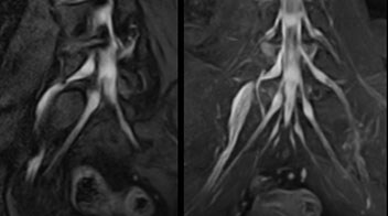
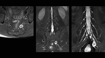
At Northern Fukushima Medical Center in Japan, excellent MRI visualization of nerves helps support confident diagnoses and informs surgical treatment decisions for patients with lower limb symptoms. MRI technologist Tanji and orthopedic surgeon Dr. Yabuki share how direct nerve visualization with the 3D NerveVIEW method adds information when diagnosing atypical herniations. The additional insights changed their way of working and benefit their patient care, as illustrated by some clinical examples.
"NerveVIEW helps us to determine the disease matching the symptoms by directly visualizing nerves"

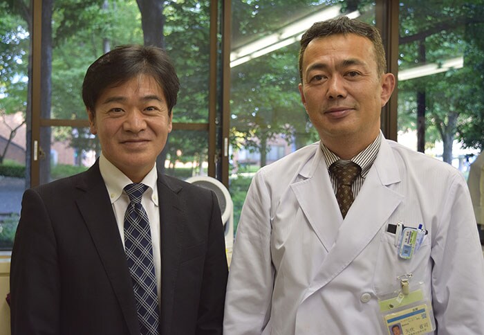
Shoji Yabuki, MD, DMSc
Shoji Yabuki, MD, DMSc is Professor at Department of Orthopaedic Surgery and Department of Pain Medicine, Fukushima Medical University School of Medicine, Fukushima City, Japan.
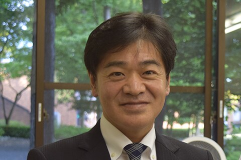
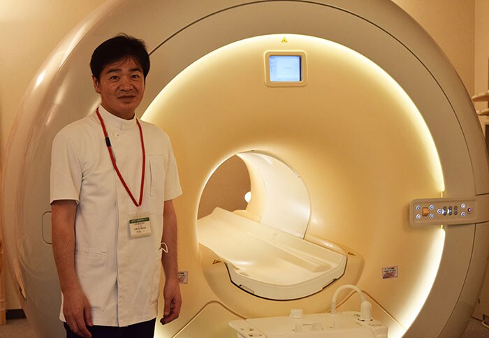
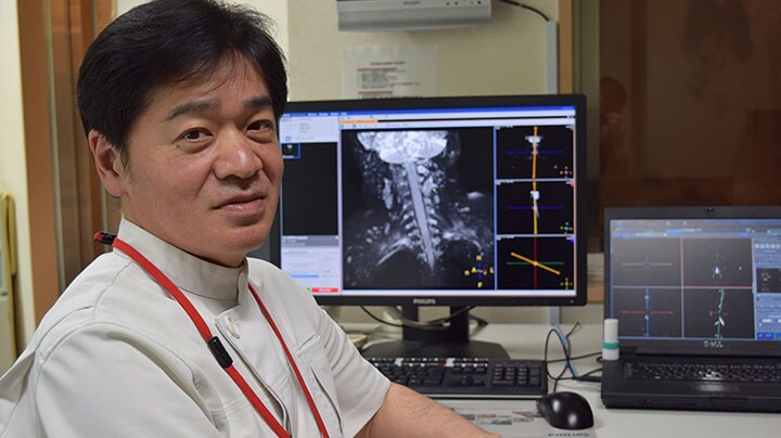
Hajime Tanji, RT
Hajime Tanji, RT is MRI technologist at Department of Radiological Technology, Imaging Center, Northern Fukushima Medical Center, Fukushima, Japan.
NerveVIEW may help when other sequences are inconclusive
“In patients with lower extremity neurological symptoms, NerveVIEW helps us to determine the disease matching the patient’s symptoms by directly visualizing the nerves. We use the sequence mainly, when there is suspicion of intraforaminal stenosis, extraforaminal stenosis or lateral disc herniation, which is often based on routine T2- and T1-weighted images. Additionally, the excellent depiction of the course of nerves makes NerveVIEW a good navigator when applying treatment such as block therapy or surgery.”
Northern Fukushima Medical Center (NFMC) Imaging Center uses the 3D NerveVIEW sequence for performing MR neurography, particularly in patients with pain and weakness in the lower limb. “It is included in about 20% of the approximately 150 lumbar spine MRI exams each month at NFMC, and can help us to determine if structures are impinging on the nerves,” says Hajime Tanji, RT, MRI technologist at NFMC.
A useful addition from surgeon’s perspective
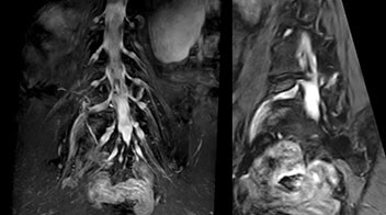
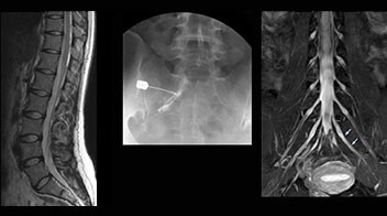
“In such case, we would then browse through axial T2-weighted MR images slice by slice and mentally reconstruct the actual situation based on both radiculography and MRI. Fortunately, NerveVIEW can now very well show nerve courses and presence of nerve compression or edema in one single image series.” “We have often seen NerveVIEW directly depict details of the nerve compression that were not observed by radiculography. Therefore, we think that with NerveVIEW we can reduce the number of invasive examinations, especially for some patients with lumbar plexus symptoms.”
“Before NerveVIEW, diagnosis by MRI alone was sometimes difficult, unless there was a strong suspicion based on clinical symptoms,” says Shoji Yabuki, MD, DMSc, Orthopedic surgeon at Fukushima Medical University School of Medicine. “This is why we routinely perform selective lumbosacral radiculography (nerve root block) and x-ray in such cases. However, radiculography can only depict nerves as far as the contrast agent reaches. When a nerve is distorted by compression, the contrast agent will not pass through this compressed area, preventing us from evaluating the full nerve compression.”
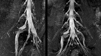
“NerveVIEW can clearly show nerve courses and presence of nerve compression"
Direct imaging of nerves aid diagnosis
The key concept in MR neurography, Dr. Yabuki stresses, is the ability to directly visualize spinal nerves, versus inferring the presence of pathology indirectly. “Before NerveVIEW, we estimated compression of the nerve by looking for the presence or absence of fat signal on other MR images,” he says.
“For example, in sagittal images, when the presence of fat is observed in the intervertebral foramen, it suggests that there is a margin around the nerve. Similarly, the absence of fat indicates that the nerve is being compressed. So, we used to deduce nerve compression indirectly. With NerveVIEW, however, we can observe the condition of the nerves directly, regardless of the presence or absence of fat. We always prefer such direct observation of anatomy over having to make an inference about it.”
Distinguishing typical from atypical herniation informs the surgeon
“Although symptoms of typical disc herniation and atypical hernia are very similar, the actual site of herniation is different. It is therefore important to characterize the nerve’s condition both inside and outside of the intervertebral foramina. “Conversely, if we see no abnormality in NerveVIEW, we can assume at least that there is no severe condition that requires surgery. Like this, it can help us avoid unnecessary surgery. NerveVIEW can have a tremendous impact in this way.”
“NerveVIEW is really useful for those cases where a nerve disorder is strongly suspected based on the clinical examination but our regular MRI images do not show any findings. These atypical herniations and spinal canal stenosis, occurring in 5% to 15% of the total lumbar herniation/stenosis cases are our main target when using NerveVIEW,” says Dr. Yabuki.
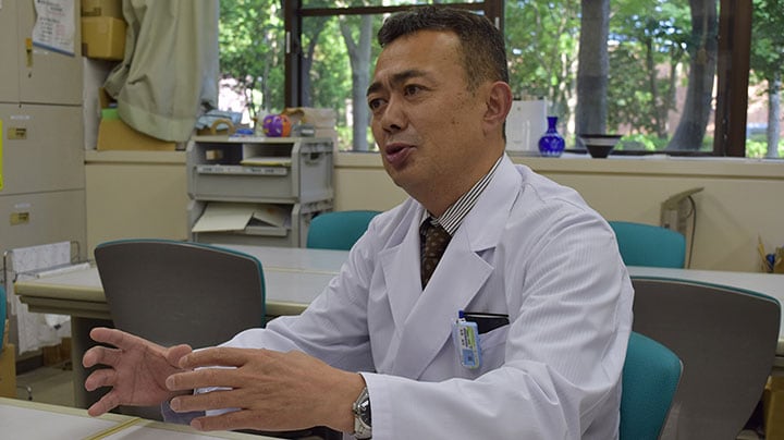
"Because NerveVIEW helps us identify the actual hernia site, it can inform selection of the surgical approach"
NFMC MRI protocol for 3D NerveVIEW in lumbar spine FOV 230 mm
Voxels 0.99 x 1.07 x 2.5 (1.25) mm
50 (100) slices
dS SENSE factor 2.0
Scan time 5:17 min.
Implementing NerveVIEW without lengthening exam time “The source images of NerveVIEW exhibit a contrast similar to STIR or fat-suppressed T2-weighted images. So, in our neurography exams we are replacing the 2D T2-weighted coronal sequence with 3D NerveVIEW. With this, we add a lot of useful information without adding scan time. This is important for patients with severe lower extremity symptoms, as they often find it difficult to maintain still during the whole MRI examination, so the exam should be as short as possible.” “We have currently implemented 3D NerveVIEW on our Achieva 3.0T dStream MRI system only. Because the 3D NerveVIEW method is based on a background signal suppression technique, we decided to use the high SNR of our 3.0T MRI system for obtaining the best possible visualization of peripheral nerves,” says Tanji. “Where NerveVIEW of the lumbar plexus is currently used as a subroutine scan for patients with strong lower limb symptoms, its use for visualization of the brachial plexus, is currently limited to special cases such as schwannomas and neuritis, usually only 1 or 2 cases per month.”
Lumbar spine MRI
examination with 3D NerveVIEW
T1W sagittal and axial
3D NerveVIEW
T2W sagittal and axial
T1W sagittal and axial
3D NerveVIEW
T1W sagittal and axial
3D NerveVIEW
Routine lumbar spine MRI
(without neurography)
T1W sagittal and axial
3D NerveVIEW
T2W sagittal and axial T1W sagittal and axial Fat suppressed T2W coronal
T1W sagittal and axial
3D NerveVIEW
"This is important for patients with severe lower extremity symptoms"
“The sequence facilitates diagnosis of lower extremity pain and informs our decision-making regarding therapy and surgery”
Building confidence with NerveVIEW
“NerveVIEW can clearly show nerve courses and presence of nerve compression. However, when multiple abnormalities are seen, it can still be hard to determine which nerve is causing the symptoms,” says Dr. Yabuki. “In our experience so far, we see abnormal findings on NerveVIEW in about 70% of elderly patients. As the pain is usually caused by only one nerve, we thus need to find the exact corresponding nerve.” “With a nerve root block, the patient's pain is improved by infiltration of local anesthesia directly around the nerve root considered to be responsible. Knowing such nerve root block findings prior to image interpretation, helps to easily recognize abnormal findings on NerveVIEW as well. In other words, without a priori knowledge, based on symptoms and/or nerve root block findings, we must be aware of the possibility of overdiagnosis.”
MR neurography attracts referrals at NFMC
The addition of the nerve-selective NerveVIEW sequence to its spine MRI protocol has given NFMC competitive advantages, according to Tanji. “Since we started including NerveVIEW routinely, the demand for lumbar spine MRI examinations has increased, especially for pre-surgical planning purposes and for patients with chronic lower extremity symptoms,” he says. “Moreover, because no other hospitals in our region are doing nerve plexus imaging yet, we often receive referrals for MR neurography studies from other hospitals even if they have an MRI scanner. Some requests come from as far as 100 km away. NerveVIEW definitely provides us a competitive advantage.” “Based on our experience, we can certainly recommend NerveVIEW to other centers,” Dr. Yabuki adds. “The sequence opens up many possibilities to facilitate the diagnosis of lower extremity pain and to inform our decision-making regarding therapy and surgery.”
