
Want to learn more about Neuro suite?
Stay up-to-date and get informed about interesting topics. Or get in touch with our sales department.
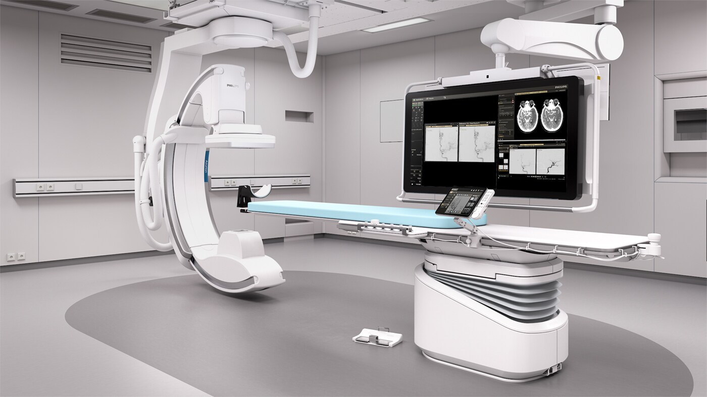
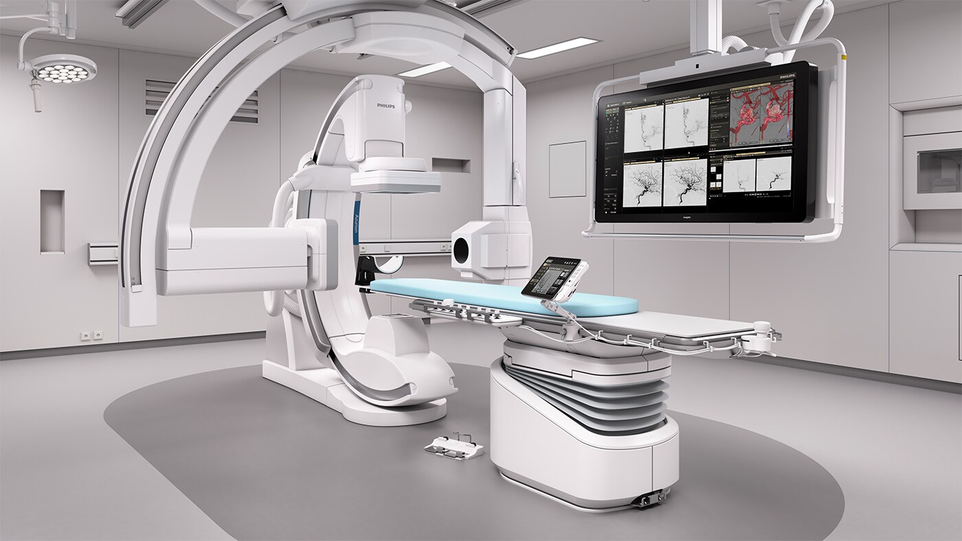
Philips Neuro suite offers a flexible portfolio of integrated technologies, services, and neuro accessories that puts you in firm control whether treating an acute stroke patient, visualizing the smallest intracranial vessels, precisely placing a flow diverter, or working slowly through a complex AVM. You can work with the confidence that comes from using sophisticated imaging technologies and neuro options that are the result of intensive research with clinical leaders and industry pioneers in neuro interventions.
Neuro suite
Clinical solutions for treating ischemic stroke
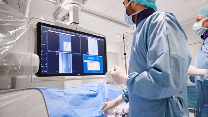
Our Neuro suite provides workflow options, dedicated interventional neuro tools, and neuro accessories to support high levels of procedural efficiency and redefine outcomes for your stroke patients. They support each step of your procedure – as you decide, guide, treat, and confirm treatment results.
Decide
The three main challenges when planning treatment are:
Comprehensive stroke diagnosis based on three XperCTs
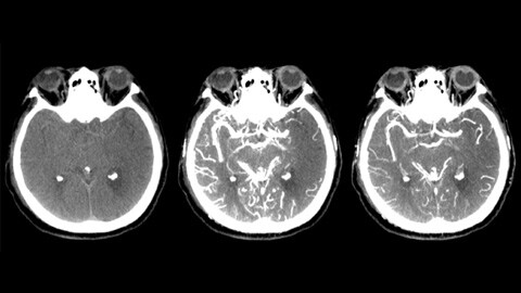
Non-contrast XperCT aids detection of early ischemic changes. Early phase XperCT helps to identify the proximal occlusion. Late phase contrast enhanced XperCT supports detection of collaterals.
Dual View to see collateral filling
Viewing early and late phase XperCT Dual volumes side by side enhances identification of penumbra and enables visualization of collateral filling.
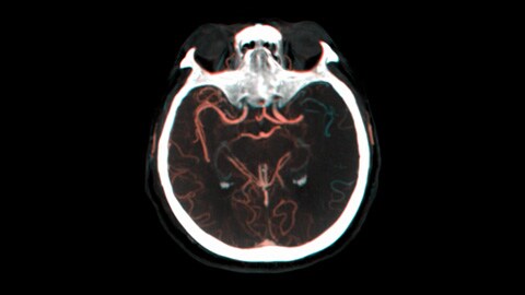
VasoCT IV to check location and length of a clot
VasoCT IV allows visualization beyond the clot with peri-procedural imaging of the distal vessel aspects in ischemic stroke. By retrograde filling, vessel structures before and after the clot become visible. The VasoCT 3D roadmap can be used to visualize clot retrieval devices.
Guide and Treat
When navigating and treating stroke pathology, clinicians need to be able to visualize the exact location of the clot and assess if and how the clot can be reached.
Maintain sharp images using 2D DSA with ClarityIQ technology
Automatic Motion Compensation during real-time DSA maintains sharp images of the vessel to support confident decision making throughout stroke procedures.
Enhance visualization of vasculature with Roadmap Pro
This advanced double contrast roadmap helps enhance visualization of overlapping vessels while balancing radiation exposure to make informed decisions about whether the clot can be reached and which route to use.
Gain anatomical references with 3D-RA and 3D Roadmap
The 3D Roadmap provides anatomical references to support precise navigation of guidewire, catheter, and device to the clot.
Confirm
After stroke treatment, there is a need to confirm if all clot material has been removed and to check for bleedings while the patient is still in the interventional lab.
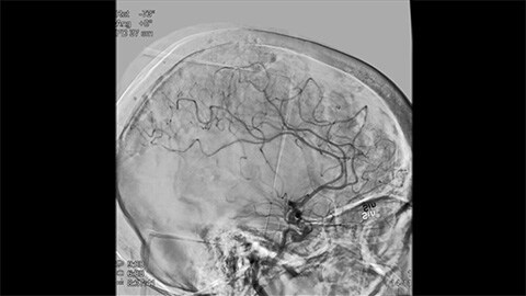
Confirm treatment success with DSA run-off
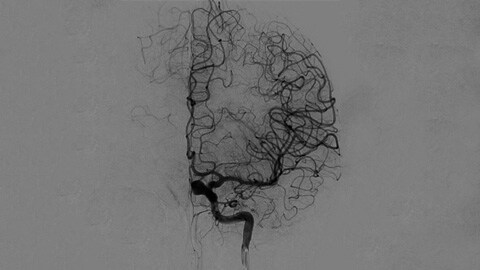
High quality DSA visualizations allow you to assess if you have retrieved the complete clot and if pieces of clot have been dispersed distally in the brain. You can check the restoration of blood flow to the penumbra and check for peri-procedure bleedings.
Peri-procedure check of bleedings with XperCT Dual
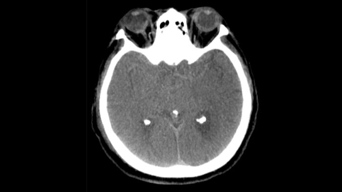
Use CT-like images in the Neuro suite to check treatment success and bleedings.
Workflow options that helps you optimize lab performance and dose management
Azurion offers a number of workflow innovations designed to help on-call staff work efficiently and easily, while maintaining a single-minded focus on the patient and manage radiation dose during acute ischemic stroke interventions.
-
-
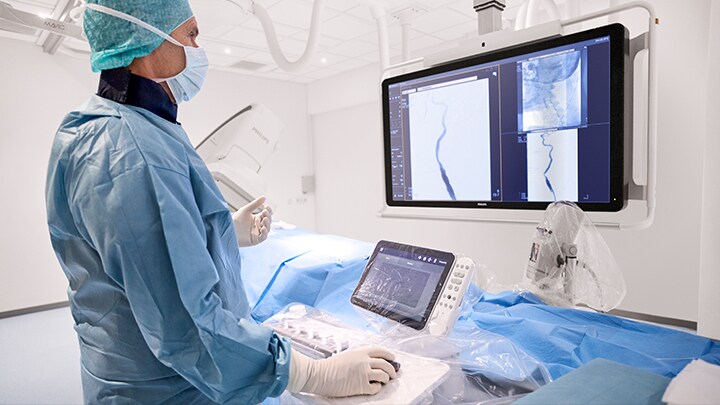
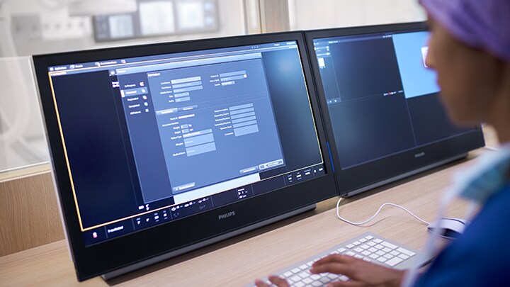
FlexVision Pro
Gives you direct access and full control of pre-operative diagnostic scans, patient information, planning tools, at table side to save time and unnecessary walking in and out of the sterile area. Touch screen module Pro allows table side control of images and applications with tablet ease.You are about to visit a Philips global content page
Continue -
Instant Parallel Working
Allows team members to work on different tasks at the same time without interrupting each other to shorten procedure times for critical stroke patients.You are about to visit a Philips global content page
Continue -
Touch Screen Module Pro
Gives you direct access and full control of pre-operative diagnostic scans, patient information, planning tools, at table side to save time and unnecessary walking in and out of the sterile area. Touch Screen Module Pro allows table side control of images and applications with tablet ease.You are about to visit a Philips global content page
Continue -
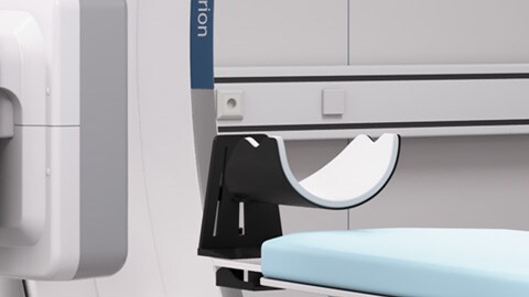
Neuro headrest
Can be used to restrain restless patients under conscious sedation to help reduce motion artefacts during the procedure.You are about to visit a Philips global content page
Continue -
ProcedureCards
ProcedureCards streamline and standardize system set-up and reduce preparation errors in acute ischemic stroke procedures. Hospital specific stroke protocols and/or checklists can be added to ProcedureCards and displayed on monitors to support consistent workflow during hectic acute situations.You are about to visit a Philips global content page
Continue
ProcedureCards
ProcedureCards streamline and standardize system set-up and help reduce preparation errors in acute ischemic stroke procedures. Hospital specific stroke protocols and/or checklists can be added to ProcedureCards and displayed on monitors to support consistent workflow during hectic acute situations.
FlexVision Pro and Touch Screen Module Pro
Gives you direct access and full control of pre-operative diagnostic scans, patient information, planning tools, at table side to save time and unnecessary walking in and out of the sterile area. Touch screen module Pro allows table side control of images and applications with tablet ease.
Instant Parallel Working
Allows team members to work on different tasks at the same time without interrupting each other to shorten procedure times for critical stroke patients.
Neuro headrest
Can be used to restrain restless patients under conscious sedation to help reduce motion artefacts during the procedure.
Integrated OR table
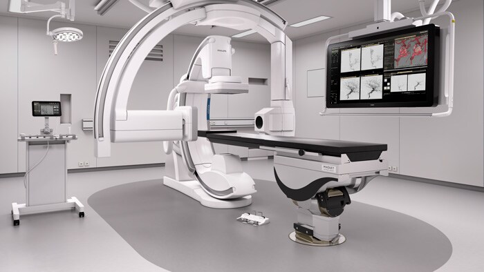
The Getinge Magnus OR table can be used for emergency and trauma care. It is synchronized with Philips X-ray systems to take advantage of advanced Philips solutions.
Case report low MAFA
Case courtesy of Prof. Laurent Spelle, Hôpital Bicêtre, Paris, France
Patient:
June 16th, 2014
December 19th, 2014
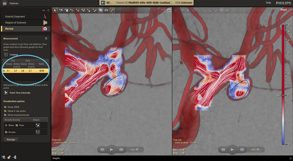
The clinical user relies on conventional DSA imaging which is the primary source of information throughout the procedure.
Case report high MAFA 1.11
Case courtesy of Dr. Vitor. Mendes Pereira, UHN, University of Toronto, Toronto, Canada
Patient:
July 19th, 2012
October 13th, 2014
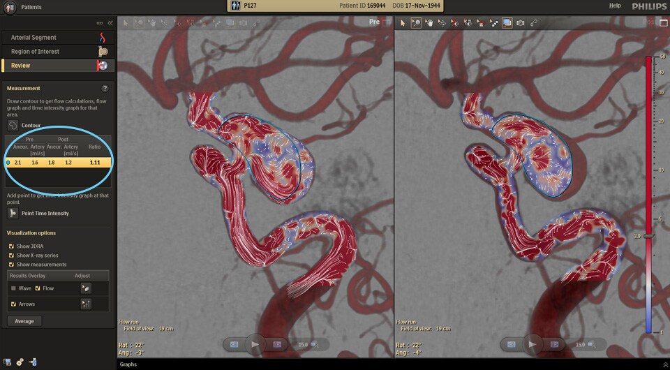
The clinical user relies on conventional DSA imaging which is the primary source of information throughout the procedure.
Decide
The three main challenges when planning cerebral aneurysm treatment are: 1) obtaining insight into tortuous vasculature, 2) accurately assessing the location, size, and neck of the aneurysm, and 3) identifying and confirming if the lesion is severe enough to require an intervention and if there is enough information to make an appropriate treatment plan.
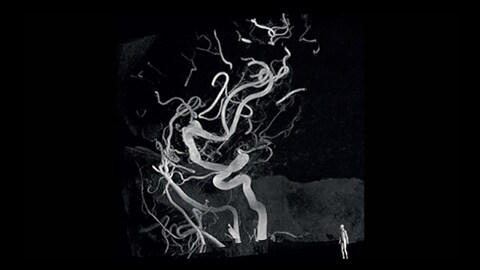
3D visualization of tortuous pathologies with 3D-RA
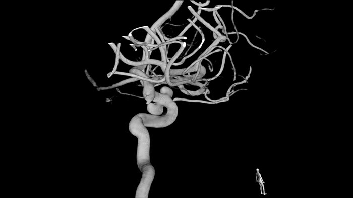
3D-RA provides a volumetric view in a few seconds to assist with assessment of location, size, neck, and severity of aneurysm for treatment planning. 3D-RA also provides high spacial resolution volumes and automatically compensates for patient movement.
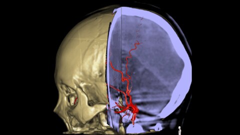
Visualize lesion boundaries and corresponding vascularization with MR-CT Roadmap
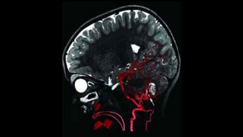
Use a previously acquired CT angio or MR angio scan and overlay it with live fluoroscopy to visualize lesion boundaries and corresponding vascularization for risk assessment. Re-using pre-acquired data helps you manage X-ray dose and contrast medium.
Guide and Treat
New technologies and devices make it more challenging than ever to efficiently navigate to the feeding vessel and accurately position devices - all while avoiding arterial dissection and spasms and minimizing contrast agent and radiation use.
Dynamic 3D image guidance through neurovascular structures
3D Roadmap enhances visualization of overlapping vessels to support precise navigation of guidewire and catheter through complex vasculature. Offers a high level of precision with real-time compensation for gantry, table, and small patient movements.
Visualize blood flow patterns with AneurysmFlow
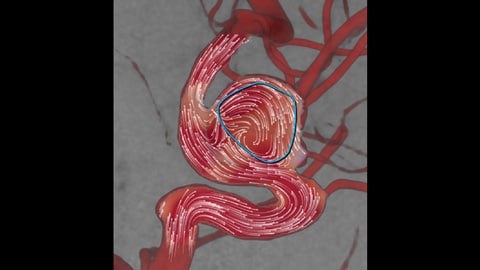
Visualize and quantify blood flow patterns in the parent vessel and aneurysm sac to obtain key information that can assist deployment of flow diverters and other embolization devices.
Support accurate guidance of devices with MR-CT Roadmap
Visualize lesion boundaries and corresponding vascularization to enhance accurate navigation through challenging pathologies, while reducing unnecessary contrast and manage X-ray dose.
Enhance visualization of cerebral vasculature with Roadmap Pro
This advanced double contrast roadmap helps enhance visualization of overlapping vessels while balancing radiation exposure. It can be customized to see advancement during coil placement.
Confirm
After aneursym treatment, check proper device placement and deployment in the context of the feeding vessel, the neck, and the sac of the aneurysm. Efficiently measure the effect of the device placed and check for possible arterial dissections while the patient is still on the table.
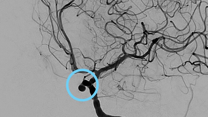
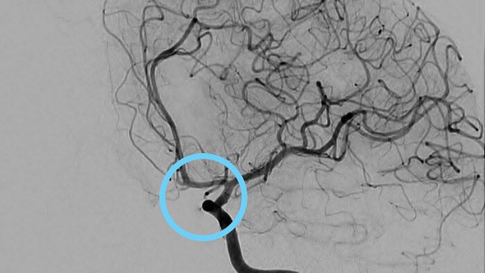
Post-treatment flow calculations with AneurysmFlow
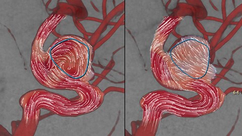
Evaluate changes in blood flow in the aneurysm pre and post, by calculating the change in Mean Aneurysm Flow Amplitude (MAFA ratio) before and after flow diverter placement.
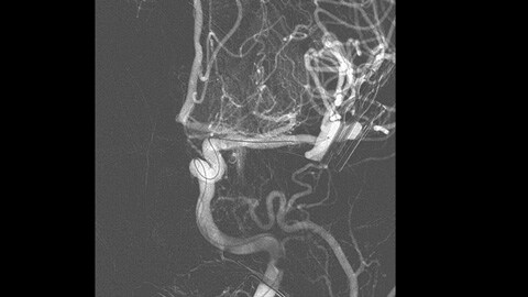
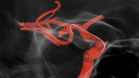
Enhance imaging of vessels and devices in the brain with VasoCT IA
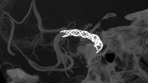
VasoCT IA is an acquisition technique that combines a high resolution XperCT with a contrast injection to enhance visualization of endovascular stents, flow diverters, and other devices and of vessel morphology down to the perforator level. It is increasingly used for follow-up of aneurysms treated with flow-diverter stents to check device positioning.
Peri-procedure check of bleedings with XperCT Dual
Use CT-like images in the Neuro suite to check treatment success and identify bleedings.
Innovative neuro interventional workflow
Our Neuro suite with Azurion offers a number of workflow innovations designed to help you simplify your workflow, shorten procedure time, and manage radiation dose during aneurysm interventions.
-
-
Neuro headrest
Can be used to help reduce motion artefacts during the procedure.You are about to visit a Philips global content page
Continue -
Full table side control with FlexVision Pro
FlexVision Pro gives you full control of all connected applications, like the CX50x Matrix ultrasound and all interventional tools at tableside to save time and unnecessary walking in and out of the sterile area.You are about to visit a Philips global content page
Continue -
Zero Dose Positioning
Helps you reduce dose by positioning the system or table on Last Image Hold so you can prepare your next run without using fluoroscopy.You are about to visit a Philips global content page
Continue -
Sharp, motionless vessels at low dose with Clarity IQ
ClarityIQ technology reduces patient dose by 75% in neuro DSA1, while maintaining equivalent image quality, compared to a system without ClarityIQ to support a broad patient population. ClarityIQ automatic motion compensation removes skull and motion artifacts which is key when placing small devices at the base of the skull.You are about to visit a Philips global content page
Continue -
Touch screen module Pro
Gives you direct access and full control of pre-operative diagnostic scans, patient information, planning tools, at table side to save time and unnecessary walking in and out of the sterile area. Touch screen module Pro allows table side control of images and applications with tablet ease.You are about to visit a Philips global content page
Continue
Sharp, motionless vessels at low dose with Clarity IQ
ClarityIQ technology reduces patient dose by 75% in neuro DSA1, while maintaining equivalent image quality, compared to a system without ClarityIQ. Its automatic motion compensation removes skull and motion artifacts which is key when placing small devices at the base of the skull.
Manage dose and simplify workflow with Zero Dose Positioning
Helps you reduce dose by positioning the system or table on Last Image Hold so you can prepare your next run without using fluoroscopy.
Full table side control with FlexVision Pro
This features helps you reduce dose by allowing you to pan the table, change table height or move the X-ray system on Last Image Hold to determine the new center position. This helps you prepare your next run without using fluoroscopy.
ProcedureCards
ProcedureCards streamline and standardize system set-up and help reduce preparation errors. Select the Aneurysm ProcedureCard and the system is set-up the way you want. Hospital specific aneurysm protocols and/or checklists can be added to ProcedureCards and displayed on monitors to support consistent workflow.
Neuro headrest
Can be used to help reduce motion artefacts during the procedure.