- Unleash the real power of MR simulation
-
Unleash the real power of MR simulation
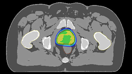
Innovative MR-only simulation pelvis lets you plan radiation therapy for male and female pelvic cancer patients with soft-tissue tumors using MRI as a single-modality solution. Within just one MR exam, MR-only simulation provides excellent soft-tissue contrast for target and OAR delineation, and CT-like density information for dose calculations. This not only extends the benefits of MRI’s outstanding soft-tissue contrast to radiotherapy planning, but it also eliminates arduous, error-prone CT-MRI registration from the process, reducing uncertainties and complexity. - Fast, automated and part of your workflow
-
Fast, automated and part of your workflow
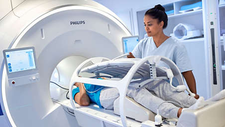
MR-only simulation pelvis is available as an option to the Ingenia MR-RT platform. It offers fast, robust scanning protocols and embedded post-processing steps to generate MRCAT (MR for Calculating ATtenuation) images with CT-like density information directly on the MR console. - Robust, consistent imaging protocol
-
Robust, consistent imaging protocol
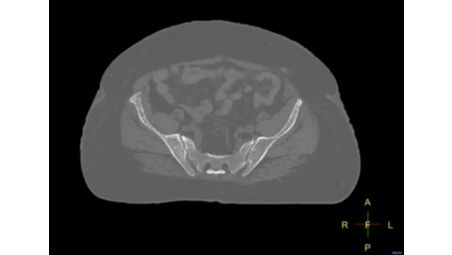
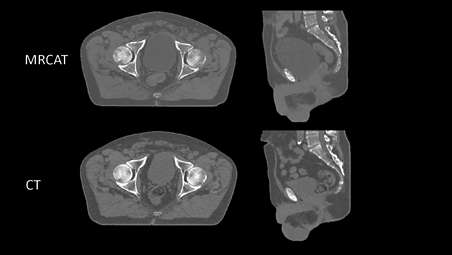
The MR-only sim imaging protocol includes a single, high-resolution, multi-contrast mDIXON sequence as the source for MRCAT generation. This scan only takes a few minutes and is standardized to deliver consistent results. The protocol is complemented with a 3D T2W scan to provide high geometric accuracy, and high-resolution image quality to support accuracy in delineation of target and critical structures. - Automatic generation of synthetic CT images
-
Automatic generation of synthetic CT images
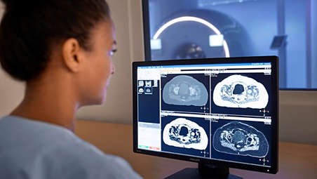
MRCAT images are generated directly on the MR using embedded image post-processing. Smart, validated algorithms that enable automatic tissue segmentation and assignment of continuous Hounsfield Units to deliver MRCAT images with CT-like density information. - Density information directly on the MR console
-
Density information directly on the MR console
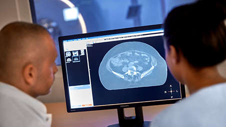
As the density information is generated directly on the MR console, the resulting data is available at the console for immediate review during the scan. The MRCAT reconstruction also automatically detects certain flaws in image acquisition, such as patient movement, and alerts the user if an immediate re-scan is necessary. This could reduce the need to call patients back for repeat exams. - MRI as primary image set in treatment planning
-
MRI as primary image set in treatment planning
The MRCAT images generated on the MR console conform to the DICOM CT standard. They can be automatically exported to your main treatment planning systems and used as the primary image dataset for dose calculations and DRR generation. - Accuracy in dose planning
-
Accuracy in dose planning
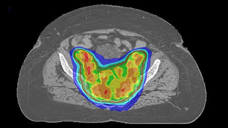
The MR-only simulation scanning protocol and MRCAT generation algorithms have been designed with the strict accuracy requirements of RT in mind. MRCAT images have high geometric accuracy¹ and validation studies have shown that MRCAT-based dose plans are robust and as accurate as CT-based plans promoting confidence in dose planning². - Patient positioning based on MR-only imaging
-
Patient positioning based on MR-only imaging
As this method eliminates CT from the process, patient positioning must also be performed as part of this workflow. The MR-based image sets with continuous Hounsfield units enable CBCT-based positioning based on soft-tissue contrast with a look and feel familiar from CT. You can also use MRCAT data to generate MR-based digitally reconstructed radiographs (DRRs) to allow for patient positioning using bony structure. - Successfully implement in your workflow
-
Successfully implement in your workflow
To successfully bring MR-only simulation into your clinical routine, we recognize the need to look beyond MR imaging – and also address steps such as patient marking, position verification, and quality assurance. We can help you successfully adopt this new simulation paradigm with our workflow descriptions and tailored training support for radiation oncology professionals. - RTdrive MR Prostate
-
RTdrive MR Prostate
MR-only simulation for pelvis includes the MR-only simulation prostate clinical application – the foundation for MR-based Auto-Contouring and RTdrive MR prostate. These innovations support the automatic contour creation of the prostate and OARs. What’s more, they allow you to generate treatment plans within 25 minutes³ including simulation imaging. This reduces the number of repetitive tasks and accelerates time-to-treatment.
Unleash the real power of MR simulation
Unleash the real power of MR simulation
Unleash the real power of MR simulation
Fast, automated and part of your workflow

Fast, automated and part of your workflow
Fast, automated and part of your workflow
Robust, consistent imaging protocol
Robust, consistent imaging protocol
Robust, consistent imaging protocol
Automatic generation of synthetic CT images
Automatic generation of synthetic CT images
Automatic generation of synthetic CT images
Density information directly on the MR console
Density information directly on the MR console
Density information directly on the MR console
MRI as primary image set in treatment planning
MRI as primary image set in treatment planning
MRI as primary image set in treatment planning
Accuracy in dose planning
Accuracy in dose planning
Accuracy in dose planning
Patient positioning based on MR-only imaging
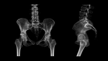
Patient positioning based on MR-only imaging
Patient positioning based on MR-only imaging
Successfully implement in your workflow
Successfully implement in your workflow
Successfully implement in your workflow
RTdrive MR Prostate
RTdrive MR Prostate
RTdrive MR Prostate
- Unleash the real power of MR simulation
- Fast, automated and part of your workflow
- Robust, consistent imaging protocol
- Automatic generation of synthetic CT images
- Unleash the real power of MR simulation
-
Unleash the real power of MR simulation
Innovative MR-only simulation pelvis lets you plan radiation therapy for male and female pelvic cancer patients with soft-tissue tumors using MRI as a single-modality solution. Within just one MR exam, MR-only simulation provides excellent soft-tissue contrast for target and OAR delineation, and CT-like density information for dose calculations. This not only extends the benefits of MRI’s outstanding soft-tissue contrast to radiotherapy planning, but it also eliminates arduous, error-prone CT-MRI registration from the process, reducing uncertainties and complexity. - Fast, automated and part of your workflow
-
Fast, automated and part of your workflow
MR-only simulation pelvis is available as an option to the Ingenia MR-RT platform. It offers fast, robust scanning protocols and embedded post-processing steps to generate MRCAT (MR for Calculating ATtenuation) images with CT-like density information directly on the MR console. - Robust, consistent imaging protocol
-
Robust, consistent imaging protocol
The MR-only sim imaging protocol includes a single, high-resolution, multi-contrast mDIXON sequence as the source for MRCAT generation. This scan only takes a few minutes and is standardized to deliver consistent results. The protocol is complemented with a 3D T2W scan to provide high geometric accuracy, and high-resolution image quality to support accuracy in delineation of target and critical structures. - Automatic generation of synthetic CT images
-
Automatic generation of synthetic CT images
MRCAT images are generated directly on the MR using embedded image post-processing. Smart, validated algorithms that enable automatic tissue segmentation and assignment of continuous Hounsfield Units to deliver MRCAT images with CT-like density information. - Density information directly on the MR console
-
Density information directly on the MR console
As the density information is generated directly on the MR console, the resulting data is available at the console for immediate review during the scan. The MRCAT reconstruction also automatically detects certain flaws in image acquisition, such as patient movement, and alerts the user if an immediate re-scan is necessary. This could reduce the need to call patients back for repeat exams. - MRI as primary image set in treatment planning
-
MRI as primary image set in treatment planning
The MRCAT images generated on the MR console conform to the DICOM CT standard. They can be automatically exported to your main treatment planning systems and used as the primary image dataset for dose calculations and DRR generation. - Accuracy in dose planning
-
Accuracy in dose planning
The MR-only simulation scanning protocol and MRCAT generation algorithms have been designed with the strict accuracy requirements of RT in mind. MRCAT images have high geometric accuracy¹ and validation studies have shown that MRCAT-based dose plans are robust and as accurate as CT-based plans promoting confidence in dose planning². - Patient positioning based on MR-only imaging
-
Patient positioning based on MR-only imaging
As this method eliminates CT from the process, patient positioning must also be performed as part of this workflow. The MR-based image sets with continuous Hounsfield units enable CBCT-based positioning based on soft-tissue contrast with a look and feel familiar from CT. You can also use MRCAT data to generate MR-based digitally reconstructed radiographs (DRRs) to allow for patient positioning using bony structure. - Successfully implement in your workflow
-
Successfully implement in your workflow
To successfully bring MR-only simulation into your clinical routine, we recognize the need to look beyond MR imaging – and also address steps such as patient marking, position verification, and quality assurance. We can help you successfully adopt this new simulation paradigm with our workflow descriptions and tailored training support for radiation oncology professionals. - RTdrive MR Prostate
-
RTdrive MR Prostate
MR-only simulation for pelvis includes the MR-only simulation prostate clinical application – the foundation for MR-based Auto-Contouring and RTdrive MR prostate. These innovations support the automatic contour creation of the prostate and OARs. What’s more, they allow you to generate treatment plans within 25 minutes³ including simulation imaging. This reduces the number of repetitive tasks and accelerates time-to-treatment.
Unleash the real power of MR simulation
Unleash the real power of MR simulation
Unleash the real power of MR simulation
Fast, automated and part of your workflow
Fast, automated and part of your workflow
Fast, automated and part of your workflow
Robust, consistent imaging protocol
Robust, consistent imaging protocol
Robust, consistent imaging protocol
Automatic generation of synthetic CT images
Automatic generation of synthetic CT images
Automatic generation of synthetic CT images
Density information directly on the MR console
Density information directly on the MR console
Density information directly on the MR console
MRI as primary image set in treatment planning
MRI as primary image set in treatment planning
MRI as primary image set in treatment planning
Accuracy in dose planning
Accuracy in dose planning
Accuracy in dose planning
Patient positioning based on MR-only imaging
Patient positioning based on MR-only imaging
Patient positioning based on MR-only imaging
Successfully implement in your workflow
Successfully implement in your workflow
Successfully implement in your workflow
RTdrive MR Prostate
RTdrive MR Prostate
RTdrive MR Prostate
Specifications
- MR-only simulation pelvis
-
MR-only simulation pelvis Anatomies - Male and female pelvis, which includes prostate, bladder, rectum, anus and cervix
Compatibility RTdrive - No
Compatibility with MR-based auto-contouring - No
Density maps - Continuous Hounsfield Units
-
- MR-only simulation prostate
-
MR-only simulation prostate Anatomies - Prostate
Compatibility RTdrive - Yes
Compatibility with MR-based auto-contouring - Yes, for prostate and prostate OARs
Density maps - Discrete, five different classifications: water, fat, spongy bone, cortical bone, and air
-
- MR-only simulation
-
MR-only simulation Compatibility - Ingenia MR-RT 1.5T and 3.0T
Imaging protocol - Dedicated ExamCard provides sequences for:
- 3D MRCAT source information (mDIXON)
- Anatomical information for delineation (T2W TSE)
MRCAT generation - Running parallel to image acquisition on the MR console, embedded post-processing generates MRCAT images using:
- Automated segmentation and tissue classification
- Automated assignment of CT-based density values
Export to treatment planning systems - MRCAT images are DICOM conform (CT) for direct export to treatment planning systems
-
- MR-only simulation pelvis
-
MR-only simulation pelvis Anatomies - Male and female pelvis, which includes prostate, bladder, rectum, anus and cervix
Compatibility RTdrive - No
-
- MR-only simulation prostate
-
MR-only simulation prostate Anatomies - Prostate
Compatibility RTdrive - Yes
-
- MR-only simulation pelvis
-
MR-only simulation pelvis Anatomies - Male and female pelvis, which includes prostate, bladder, rectum, anus and cervix
Compatibility RTdrive - No
Compatibility with MR-based auto-contouring - No
Density maps - Continuous Hounsfield Units
-
- MR-only simulation prostate
-
MR-only simulation prostate Anatomies - Prostate
Compatibility RTdrive - Yes
Compatibility with MR-based auto-contouring - Yes, for prostate and prostate OARs
Density maps - Discrete, five different classifications: water, fat, spongy bone, cortical bone, and air
-
- MR-only simulation
-
MR-only simulation Compatibility - Ingenia MR-RT 1.5T and 3.0T
Imaging protocol - Dedicated ExamCard provides sequences for:
- 3D MRCAT source information (mDIXON)
- Anatomical information for delineation (T2W TSE)
MRCAT generation - Running parallel to image acquisition on the MR console, embedded post-processing generates MRCAT images using:
- Automated segmentation and tissue classification
- Automated assignment of CT-based density values
Export to treatment planning systems - MRCAT images are DICOM conform (CT) for direct export to treatment planning systems
-
Related products
Alternative products
-
Big Bore RT
- Advance confidence in clinical diagnosis and treatment planning
- Accelerate time to treatment through intuitive workflow tools
- Enhance patient/staff satisfaction by creating positive experiences
- Maximize value with cross purpose oncology/radiology configuration
View product
-
Ingenia Ambition/Elition MR-RT
- Drive the precision of radiation therapy
- A superb MRI platform for radiation oncology
- Position with precision
- Set up easily and flexibly
View product


-
Big Bore RT
Big Bore RT is designed as a CT simulator to enhance clinical confidence, accelerate time to treat and maximize value of its investment without compromising on patient experience – four dimensions that are essential towards excellent care.
View product
-
Ingenia Ambition/Elition MR-RT
The state-of-the-art Ingenia MR-RT platform featuring Ambition 1.5T and Elition 3.0T MR systems meets specific RT needs by providing high-quality MR images acquired in the treatment position. Smoothly integrate MRI through a comprehensive solution that considers your whole workflow, even for MR-only radiotherapy.
View product
- 1. Accuracy means: MRCAT provides around 1 mm total geometric accuracy of image data in less than 20 cm Diameter Spherical Volume (DSV). MRCAT provides below 2 mm total geometric accuracy of image data in more than 40 cm Diameter Spherical Volume (DSV)*. * Limited to 32 cm in z-direction in more than 95% of the points within the volume
- 2. Equivalent means: the simulated dose based on MRCAT images does not differ in 95% of the pelvic cancer patients (Gamma analysis criterion 3%/3mm realized in 99% of voxels within the PTV or exceeding 75% of the maximum dose) when compared with CT-based plan for EBRT.
- 3. Tested in a non-clinical environment with single Pinnacle user and a 5-beam IMRT plan. Excluding time for optional manual adjustments.