- nSIGHT Imaging
-
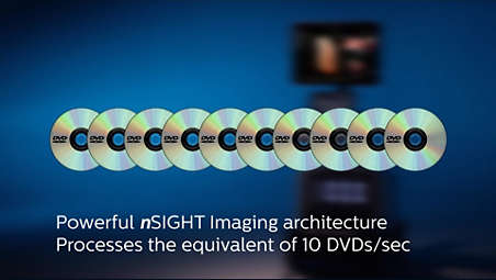
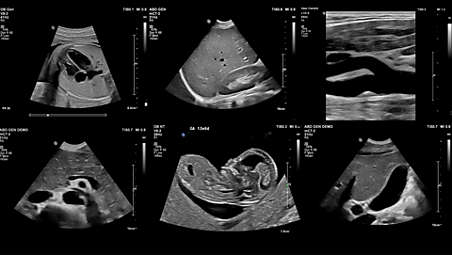
nSIGHT Imaging
Far surpasses conventional ultrasound performance to reach new levels of definition and clarity. Incorporating a custom multi-stage precision beamformer along with massiveparallel processing, this proprietary architecture captures an enormous amount of acoustic data from each transmit operation and performs digital beam reconstruction along with mathematically optimized focal processing. This creates extraordinary real-time images with exceptional frame rate, uniformity and penetration. - FlexVue with Orthogonal View
-
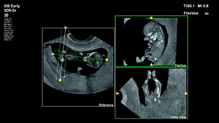
FlexVue with Orthogonal View
95% of OB/GYN users surveyed* feel, FlexVue and Orthogonal View, improves their workflow and 85% say it enhances their diagnostic confidence as well. - MFI HD
-
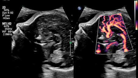
MFI HD
85% of OB/GYN users surveyed*, feel MFI HD, with its ultrasensitive blood flow imaging, enhances their diagnostic confidence. - 24" HD MAX display
-
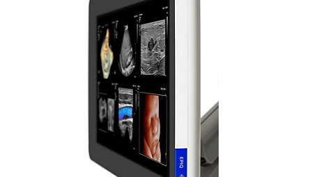
24" HD MAX display
This new immersive 24” display monitor offers the ultimate ultrasound visualization experience, with an ultra-wide color gamut of 10-bit color depth that uses billions of colors for accurate color reproduction. In addition, it provides high-contrast dynamic range and enhanced black levels for subtle delineation of grayscale values. HD MAX features superb off-angle viewing for visualization of clinical images throughout the scanning room. - XRES Pro, the next-generation image processing
-
XRES Pro, the next-generation image processing
At real-time frame rates, XRES Pro uses multi-parametric precision filters that subdivide image elements, analyze this data and then apply advanced algorithms to sharpen borders and interfaces and provide superb tissue conspicuity. XRES Pro also offers enhanced assessment of plaque morphology. XRES Pro allows you full adjustability to match the level of enhancement to clinical imaging requirements for elevated diagnostic confidence with virtually all patients. - MicroFlow Imaging
-
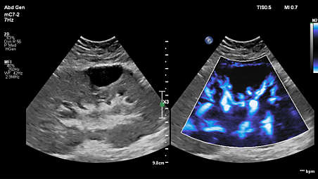
MicroFlow Imaging
Designed to detect slow and weak blood flow anatomy in tissue. This proprietary approach overcomes many of the barriers associated with conventional methods to detect small vessel blood flow with high resolution and minimal artifacts. MicroFlow Imaging maintains high frame rate and 2D image quality while applying advanced artifact reduction techniques to reveal small vessel anatomy - PureWave & xMATRIX technology
-
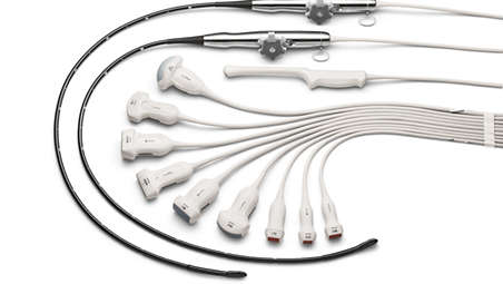
PureWave & xMATRIX transducer technology
The power of PureWave for exceptional imaging even on technically difficult patients. PureWave crystal technology represents the biggest breakthrough in piezoelectric transducer material in 40 years. The pure, uniform crystals of PureWave have virtually perfect uniformity for greater bandwidth and twice the efficiency of conventional ceramic materials. - xMATRIX transducers, powerful and versatile
-
xMATRIX transducers, powerful and versatile
No other premium ultrasound solution can run xMATRIX, the comprehensive suite of the world’s most innovative ultrasound transducers. Achieve ultra-thin 2D slices. Use Live xPlane imaging to create two full-resolution planes simultaneously, allowing you to capture twice as much clinical information in the same amount of time. Acquire near-isovoxel resolution to reveal images from any plane within the volume. - Anatomically Intelligent ultrasound - machine intelligence for faster more reproducible analysis
-
Anatomically Intelligent ultrasound - machine intelligence for faster more reproducible analysis
At the heart of the powerful EPIQ Elite architecture is our Philips exclusive Anatomical Intelligence Ultrasound (AIUS), designed to elevate the ultrasound system from a passive to an actively adaptive device. With advanced organ modeling, image slicing, and proven quantification, exams are easy to perform, more reproducible, and deliver new levels of clinical information. Some examples of our AIUS capabilities include HeartModel, AI Breast and Auto-registration of image fusion and navigation. - Image Fusion and Navigation-Easy to use modality fusion and interventional guidance
-
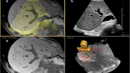
Image Fusion and Navigation-Easy to use modality fusion and interventional guidance
Make confident decisions even in challenging diagnostic cases with fully integrated fusion capabilities that feature streamlined workflows to allow clinicians to achieve fast and effective fusion of CT/MR/PET with live ultrasound. By combining imaging modalities directly on the ultrasound system, you now have access to an even more powerful diagnostic tool with advanced visualization allowing for fast clinical decisions. Expand fusion and navigation capabilities through a range of transducers across applications, including the X6-1 xMATRIX, C5-1, C9-2, eL18-4, L12-5, C10-4ec, S5-1 and the new mC7-2. - Powerful system security - protecting sensitive patient data
-
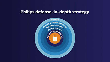
Powerful system security - protecting sensitive patient data
Hospitals and healthcare organizations are spending more to protect their systems and patient data from cyber-attacks. That is why healthcare cybersecurity spending will exceed $65 billion over the next five years. Ultrasound devices are highly mobile and can exist in a wired or wireless environment. As a result, Philips has made security a high priority for ultrasound systems. The EPIQ Elite platform is built on Window 10 OS and features a powerful defense-in-depth principle and deliver an outstanding set of data security features comprising of five core layers. - Elastography
-
Elastography
The EPIQ Elite platform supports both strain and shear wave imaging methods of elastography. Highly sensitive strain imaging can be used to rapidly assess relative tissue stiffness values across a variety of applications. ElastQ Imaging methods of shear wave elastography use a unique pulsing scheme to generate and detect the propagation speed of shear waves, providing a quantitative display and measurement of tissue stiffness. ElastQ Imaging also provides a confidence map display to assist you in obtaining measurements from areas with the highest shear wave quality.
nSIGHT Imaging
nSIGHT Imaging
nSIGHT Imaging
FlexVue with Orthogonal View
FlexVue with Orthogonal View
FlexVue with Orthogonal View
MFI HD
MFI HD
MFI HD
24" HD MAX display
24" HD MAX display
24" HD MAX display
XRES Pro, the next-generation image processing
XRES Pro, the next-generation image processing
XRES Pro, the next-generation image processing
MicroFlow Imaging
MicroFlow Imaging
MicroFlow Imaging
PureWave & xMATRIX transducer technology
PureWave & xMATRIX transducer technology
PureWave & xMATRIX transducer technology
xMATRIX transducers, powerful and versatile

xMATRIX transducers, powerful and versatile
xMATRIX transducers, powerful and versatile
Anatomically Intelligent ultrasound - machine intelligence for faster more reproducible analysis
Anatomically Intelligent ultrasound - machine intelligence for faster more reproducible analysis
Anatomically Intelligent ultrasound - machine intelligence for faster more reproducible analysis
Image Fusion and Navigation-Easy to use modality fusion and interventional guidance
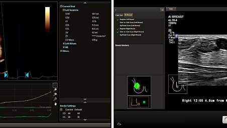
Image Fusion and Navigation-Easy to use modality fusion and interventional guidance
Image Fusion and Navigation-Easy to use modality fusion and interventional guidance
Powerful system security - protecting sensitive patient data
Powerful system security - protecting sensitive patient data
Powerful system security - protecting sensitive patient data
Elastography
Elastography
- nSIGHT Imaging
- FlexVue with Orthogonal View
- MFI HD
- 24" HD MAX display
- nSIGHT Imaging
-
nSIGHT Imaging
Far surpasses conventional ultrasound performance to reach new levels of definition and clarity. Incorporating a custom multi-stage precision beamformer along with massiveparallel processing, this proprietary architecture captures an enormous amount of acoustic data from each transmit operation and performs digital beam reconstruction along with mathematically optimized focal processing. This creates extraordinary real-time images with exceptional frame rate, uniformity and penetration. - FlexVue with Orthogonal View
-
FlexVue with Orthogonal View
95% of OB/GYN users surveyed* feel, FlexVue and Orthogonal View, improves their workflow and 85% say it enhances their diagnostic confidence as well. - MFI HD
-
MFI HD
85% of OB/GYN users surveyed*, feel MFI HD, with its ultrasensitive blood flow imaging, enhances their diagnostic confidence. - 24" HD MAX display
-
24" HD MAX display
This new immersive 24” display monitor offers the ultimate ultrasound visualization experience, with an ultra-wide color gamut of 10-bit color depth that uses billions of colors for accurate color reproduction. In addition, it provides high-contrast dynamic range and enhanced black levels for subtle delineation of grayscale values. HD MAX features superb off-angle viewing for visualization of clinical images throughout the scanning room. - XRES Pro, the next-generation image processing
-
XRES Pro, the next-generation image processing
At real-time frame rates, XRES Pro uses multi-parametric precision filters that subdivide image elements, analyze this data and then apply advanced algorithms to sharpen borders and interfaces and provide superb tissue conspicuity. XRES Pro also offers enhanced assessment of plaque morphology. XRES Pro allows you full adjustability to match the level of enhancement to clinical imaging requirements for elevated diagnostic confidence with virtually all patients. - MicroFlow Imaging
-
MicroFlow Imaging
Designed to detect slow and weak blood flow anatomy in tissue. This proprietary approach overcomes many of the barriers associated with conventional methods to detect small vessel blood flow with high resolution and minimal artifacts. MicroFlow Imaging maintains high frame rate and 2D image quality while applying advanced artifact reduction techniques to reveal small vessel anatomy - PureWave & xMATRIX technology
-
PureWave & xMATRIX transducer technology
The power of PureWave for exceptional imaging even on technically difficult patients. PureWave crystal technology represents the biggest breakthrough in piezoelectric transducer material in 40 years. The pure, uniform crystals of PureWave have virtually perfect uniformity for greater bandwidth and twice the efficiency of conventional ceramic materials. - xMATRIX transducers, powerful and versatile
-
xMATRIX transducers, powerful and versatile
No other premium ultrasound solution can run xMATRIX, the comprehensive suite of the world’s most innovative ultrasound transducers. Achieve ultra-thin 2D slices. Use Live xPlane imaging to create two full-resolution planes simultaneously, allowing you to capture twice as much clinical information in the same amount of time. Acquire near-isovoxel resolution to reveal images from any plane within the volume. - Anatomically Intelligent ultrasound - machine intelligence for faster more reproducible analysis
-
Anatomically Intelligent ultrasound - machine intelligence for faster more reproducible analysis
At the heart of the powerful EPIQ Elite architecture is our Philips exclusive Anatomical Intelligence Ultrasound (AIUS), designed to elevate the ultrasound system from a passive to an actively adaptive device. With advanced organ modeling, image slicing, and proven quantification, exams are easy to perform, more reproducible, and deliver new levels of clinical information. Some examples of our AIUS capabilities include HeartModel, AI Breast and Auto-registration of image fusion and navigation. - Image Fusion and Navigation-Easy to use modality fusion and interventional guidance
-
Image Fusion and Navigation-Easy to use modality fusion and interventional guidance
Make confident decisions even in challenging diagnostic cases with fully integrated fusion capabilities that feature streamlined workflows to allow clinicians to achieve fast and effective fusion of CT/MR/PET with live ultrasound. By combining imaging modalities directly on the ultrasound system, you now have access to an even more powerful diagnostic tool with advanced visualization allowing for fast clinical decisions. Expand fusion and navigation capabilities through a range of transducers across applications, including the X6-1 xMATRIX, C5-1, C9-2, eL18-4, L12-5, C10-4ec, S5-1 and the new mC7-2. - Powerful system security - protecting sensitive patient data
-
Powerful system security - protecting sensitive patient data
Hospitals and healthcare organizations are spending more to protect their systems and patient data from cyber-attacks. That is why healthcare cybersecurity spending will exceed $65 billion over the next five years. Ultrasound devices are highly mobile and can exist in a wired or wireless environment. As a result, Philips has made security a high priority for ultrasound systems. The EPIQ Elite platform is built on Window 10 OS and features a powerful defense-in-depth principle and deliver an outstanding set of data security features comprising of five core layers. - Elastography
-
Elastography
The EPIQ Elite platform supports both strain and shear wave imaging methods of elastography. Highly sensitive strain imaging can be used to rapidly assess relative tissue stiffness values across a variety of applications. ElastQ Imaging methods of shear wave elastography use a unique pulsing scheme to generate and detect the propagation speed of shear waves, providing a quantitative display and measurement of tissue stiffness. ElastQ Imaging also provides a confidence map display to assist you in obtaining measurements from areas with the highest shear wave quality.
nSIGHT Imaging
nSIGHT Imaging
nSIGHT Imaging
FlexVue with Orthogonal View
FlexVue with Orthogonal View
FlexVue with Orthogonal View
MFI HD
MFI HD
MFI HD
24" HD MAX display
24" HD MAX display
24" HD MAX display
XRES Pro, the next-generation image processing
XRES Pro, the next-generation image processing
XRES Pro, the next-generation image processing
MicroFlow Imaging
MicroFlow Imaging
MicroFlow Imaging
PureWave & xMATRIX transducer technology
PureWave & xMATRIX transducer technology
PureWave & xMATRIX transducer technology
xMATRIX transducers, powerful and versatile
xMATRIX transducers, powerful and versatile
xMATRIX transducers, powerful and versatile
Anatomically Intelligent ultrasound - machine intelligence for faster more reproducible analysis
Anatomically Intelligent ultrasound - machine intelligence for faster more reproducible analysis
Anatomically Intelligent ultrasound - machine intelligence for faster more reproducible analysis
Image Fusion and Navigation-Easy to use modality fusion and interventional guidance
Image Fusion and Navigation-Easy to use modality fusion and interventional guidance
Image Fusion and Navigation-Easy to use modality fusion and interventional guidance
Powerful system security - protecting sensitive patient data
Powerful system security - protecting sensitive patient data
Powerful system security - protecting sensitive patient data
Elastography
Elastography
Specifications
- Common Specifications
-
Common Specifications Width - 60.6 cm/ 23.9 in
Height - 146-171.5 cm/ 57.5-67.5 in
Depth - 109.2 cm/ 43 in
Weight - 104.3 kg/ 230 lb without peripheral devices
-
- Control panel
-
Control panel Monitor size - 24 inch / 60.96 cm HD display
-
- Common Specifications
-
Common Specifications Width - 60.6 cm/ 23.9 in
Height - 146-171.5 cm/ 57.5-67.5 in
-
- Control panel
-
Control panel Monitor size - 24 inch / 60.96 cm HD display
-
- Common Specifications
-
Common Specifications Width - 60.6 cm/ 23.9 in
Height - 146-171.5 cm/ 57.5-67.5 in
Depth - 109.2 cm/ 43 in
Weight - 104.3 kg/ 230 lb without peripheral devices
-
- Control panel
-
Control panel Monitor size - 24 inch / 60.96 cm HD display
-
- *based on a sample size of 20 users